

Where and how does the pain appear?
Superficial or deep, pinpoint or widespread, pulling, stabbing, burning, pressing or electric: the precise quality determines which levels are assessed first.
Pain & functional symptoms
The location of pain is the starting point, not automatically the complete explanation. In Applied Neurofunction, skin, muscle, fascia, tendon, joint, nerve, blood vessels and processing are assessed individually, suitable inputs are identified and the original pain or movement response is re-tested straight away.
How functional assessment works →About the images: The scene images on this page are AI-generated illustrations. They do not show real clients or document actual sessions in the practice.
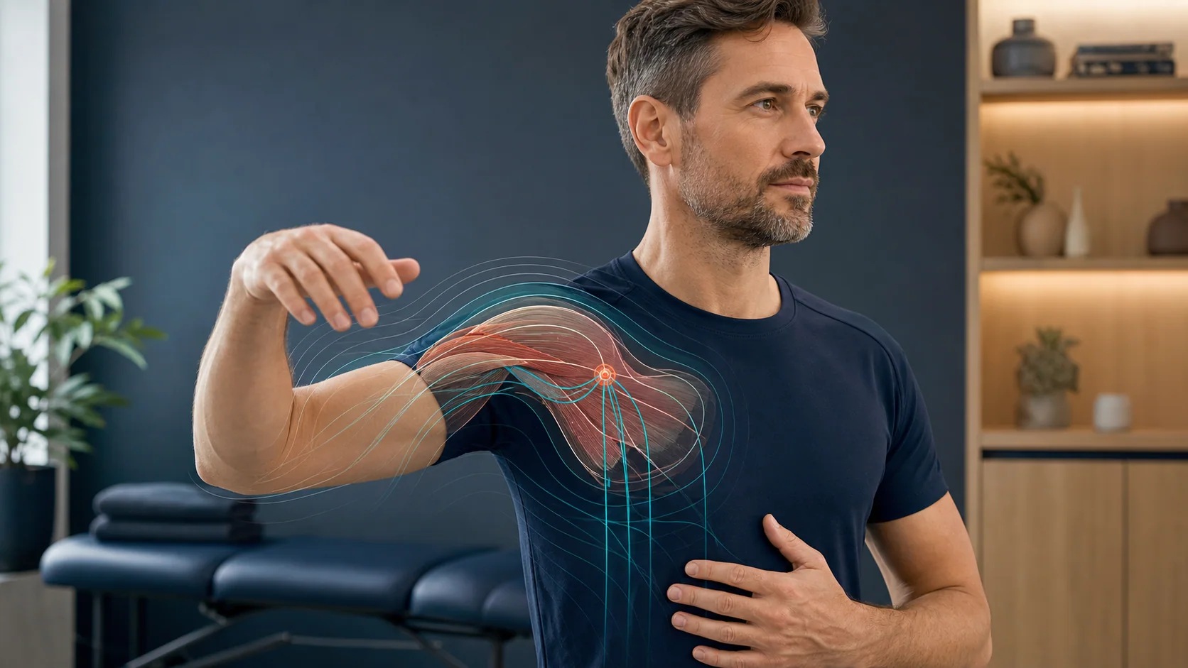
From a symptom to a functional map
“My shoulder hurts” or “my knee is painful” initially describes the response. A targeted approach asks more precise questions: which movement triggers it? Which layer reacts? Which direction, load, position or sensory information changes it?
Superficial or deep, pinpoint or widespread, pulling, stabbing, burning, pressing or electric: the precise quality determines which levels are assessed first.
Joint angle, direction of movement, speed, muscle contraction, stretch, load and number of repetitions are varied separately. This shows which variable makes the greatest difference.
Touch, pressure, tissue tension, joint position, peripheral nerves and supply pathways provide different information. Each level may show a finding on its own or only react in combination.
Vision, balance, breathing, stress state, protective tension and movement planning influence how safely a movement is evaluated and organised.
A tissue map rather than a broad label
Each level is assessed with appropriate inputs and movement conditions. This narrows the search from a general region to the structure, direction and function that are currently relevant.
Light touch, pressure, direction of pull, temperature and local mobility are considered separately. A superficial input may immediately change a movement or pain response.
Tissue tension, displacement, direction and the gliding of neighbouring layers are assessed separately. What matters is whether a particular direction of pull improves the initial test.
Contraction, muscle length, joint angle, strength and the function of muscle spindles, tendons and fascia are assessed individually rather than combined into a single “muscle test”.
Active and passive movement, end position, axis of movement, joint position and sensory feedback help show whether the difficulty arises from the joint itself or from its control.
Measured pressure, tapping and direction can be assessed separately. This level becomes particularly relevant when a very local, deep point of discomfort stands out.
Sensation, strength, nerve course and head, shoulder, arm or leg position are considered together. Reducing tension, gliding and increasing mechanical tension are different assessment conditions.
Position- and direction-dependent responses, differences between sides and relationships with inflow and outflow are assessed and compared with the original response.
Somatosensation, vision, balance, the brainstem, cerebellum, breathing and autonomic state are included when the local structure alone does not fully explain the pattern.
Back, neck, shoulder, hip, knee & foot
A painful movement involves joint position, tissue loading, sensory feedback, neural pathways and motor organisation. Only by distinguishing these can we identify where the most effective change is possible.
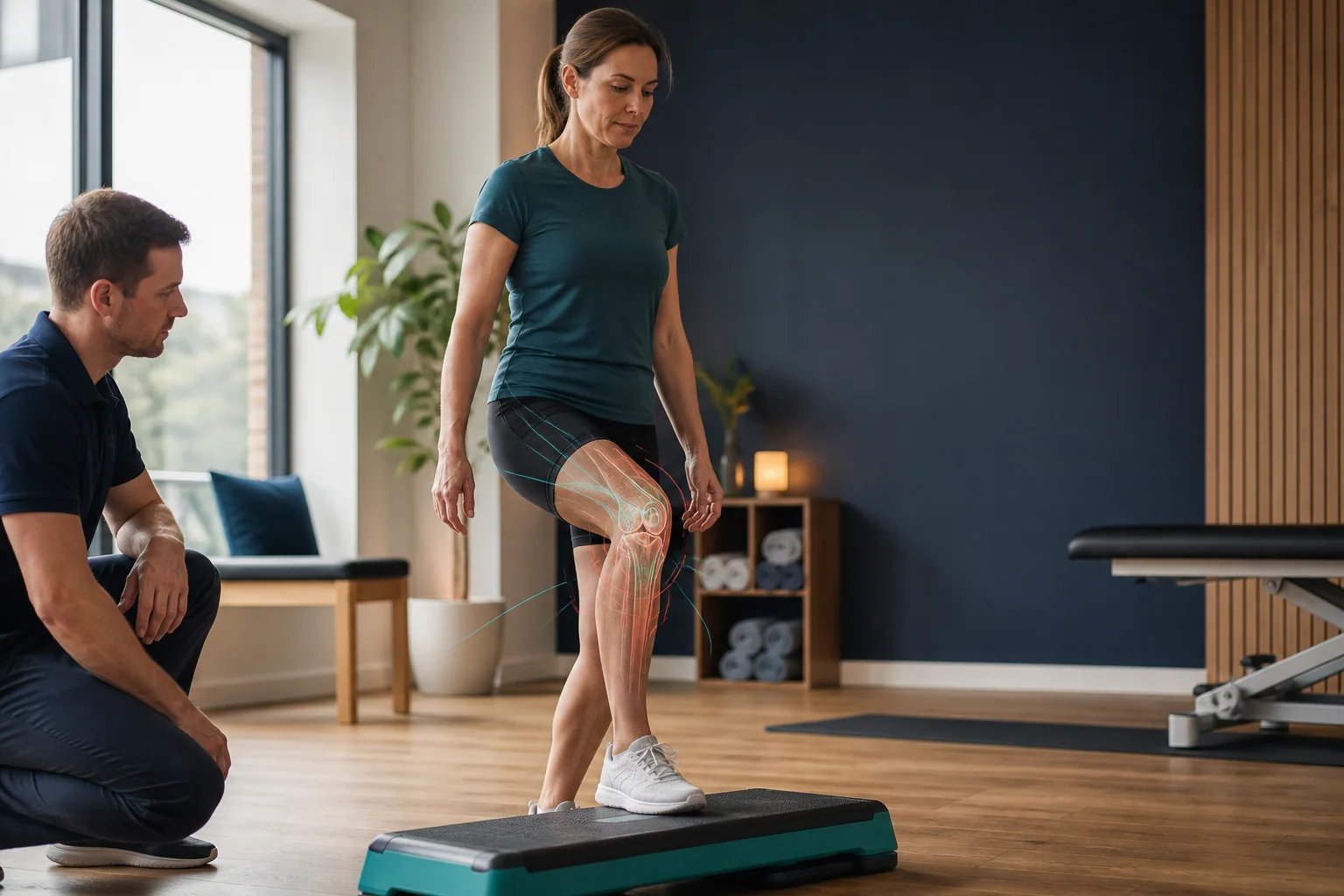
Whether your arm hurts when reaching into a cupboard, your knee on stairs or your back when straightening up: that exact everyday movement is recorded as the baseline first. It is then broken down into its components, rather than simply repeated.
Direction, angle, speed, load, onset of pain and visible compensation are defined so they can be reproduced.
Skin, fascia, muscle, tendon, joint, bone, nerve and blood vessels are assessed using inputs and positions suited to each.
If a functional difficulty remains unresolved, joint position, spinal stability, balance, vision, breathing and other relevant relationships are explored.
Each successful individual input is integrated. The same movement is then repeated with identical angle and speed and a comparable load.
When symptoms do not fit a simple category
Functional symptoms may depend on position, vary or be difficult to locate. In these cases, it is useful to ask which pathway, layer or processing condition reproducibly changes the symptom.
Distribution, sensation, strength, arm or leg position, head posture, nerve course and supply are compared systematically.
The quality of the symptom is related to skin inputs, peripheral nerve mechanics, local tissue tension and central sensory processing.
Load, sleep, stress, breathing, body position, sensory surroundings and day-to-day condition can change which function reaches its limit first.
Alongside intensity, movement quality, stability, strength, endurance and the specific everyday task are recorded as equally important re-test measures.
Three specific assessment routes
These examples show how musculoskeletal pain, tingling and migraine are broken down into different functions. The individual's starting pattern is always central.
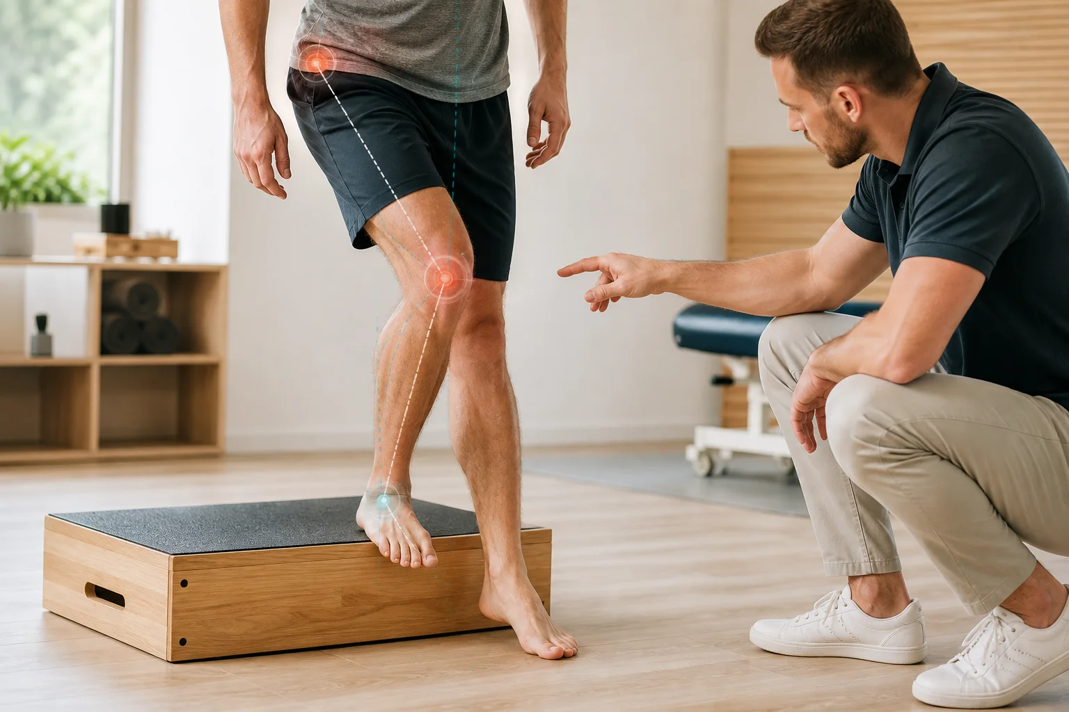
Scenario 1 · Musculoskeletal knee pain
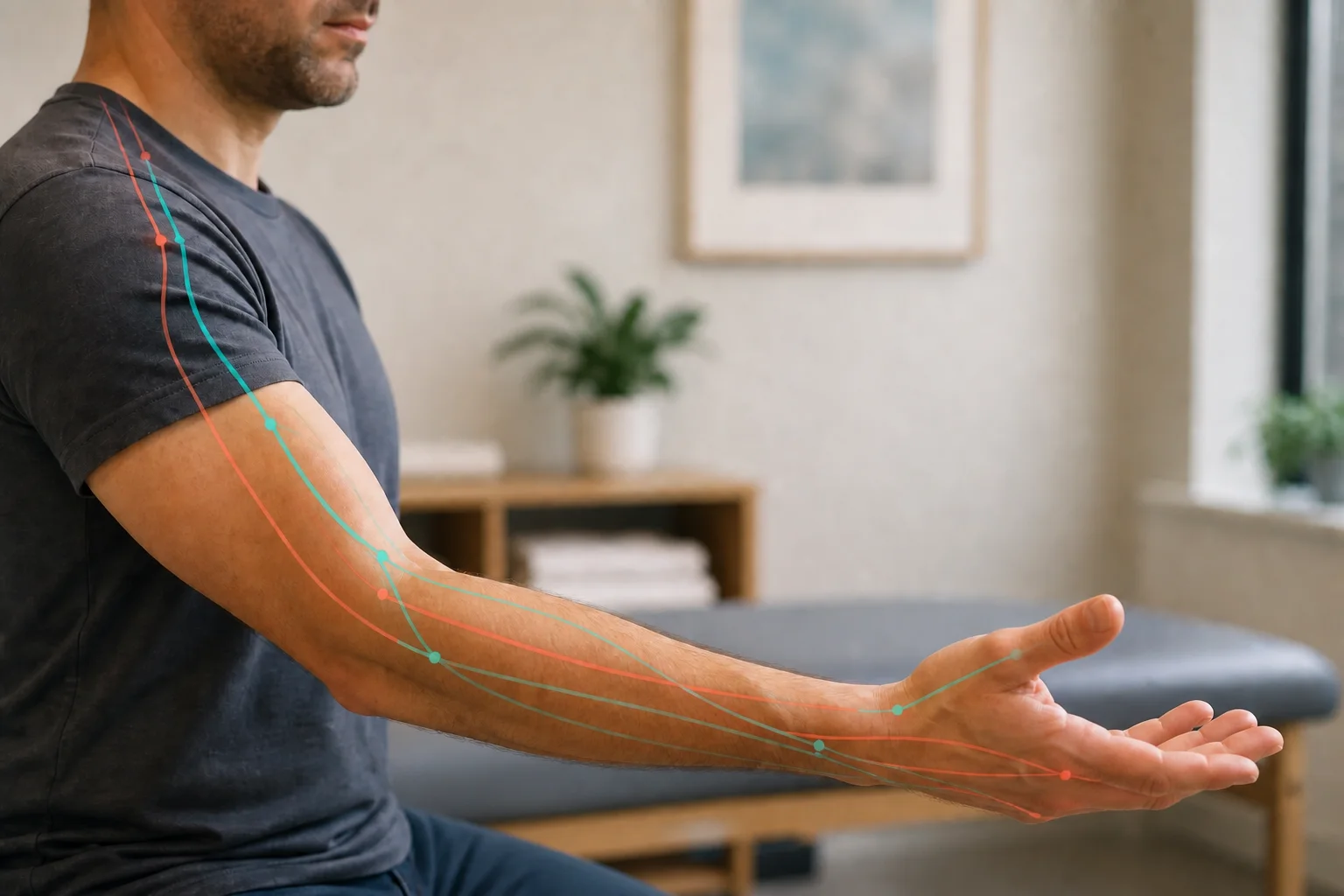
Scenario 2 · Tingling in the hand and arm
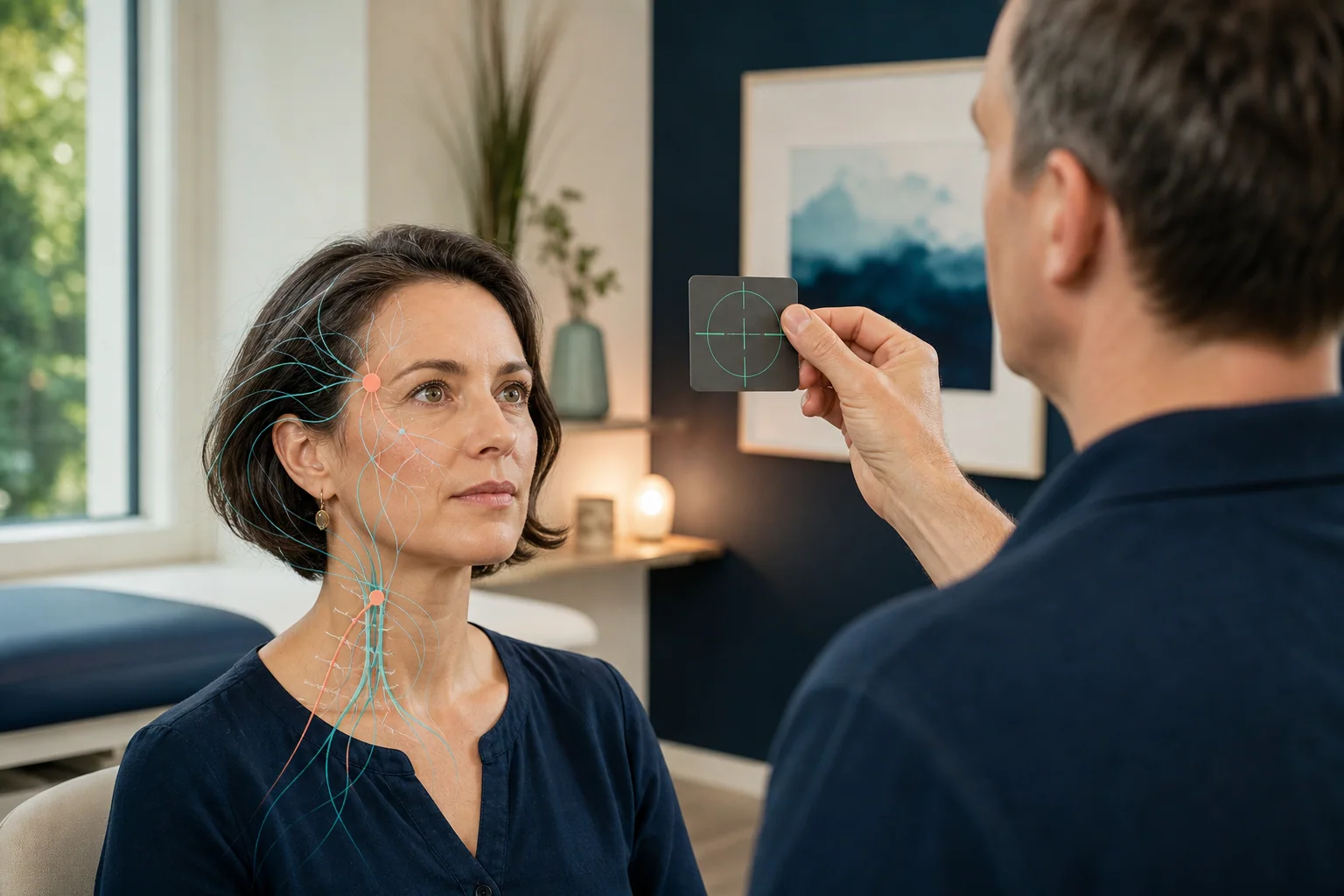
Scenario 3 · Migraine with visual and autonomic involvement
Breaking down migraine functionally
Migraine may involve pain, vision, balance, nausea, the neck, energy and autonomic regulation at the same time. The functional assessment follows this overall pattern.
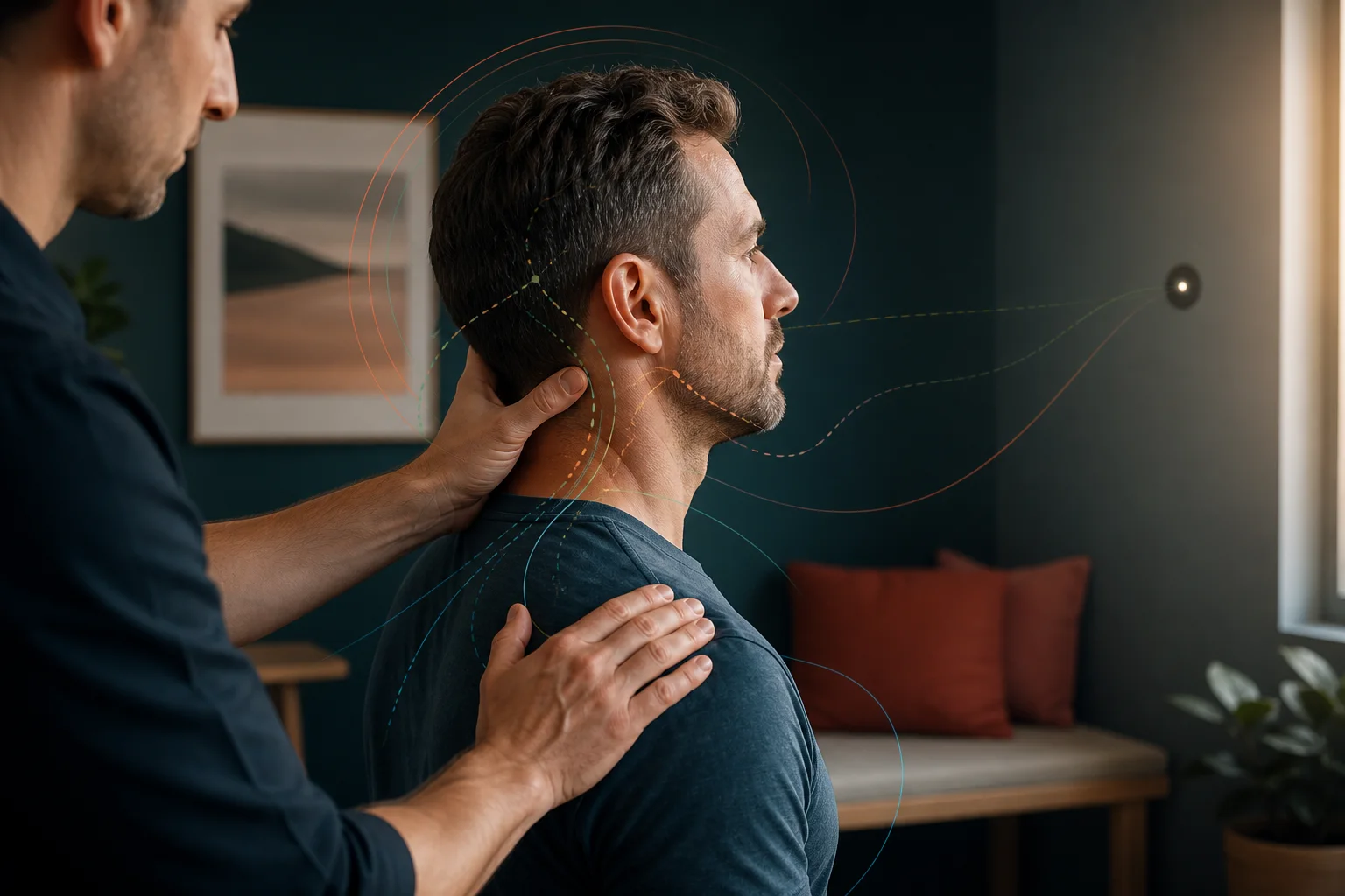
First, we understand the sequence of the attack: early signs, aura, the pain phase itself and recovery may reveal different functions. The history therefore includes not only frequency and pain intensity, but also light, sounds, movement, nausea, neck stiffness, sleep, stress, menstrual cycle and meal timing.
The next step does not follow a fixed migraine programme. Visual, trigeminal, cervical, vestibular, autonomic and cranial functions are assessed individually. Findings are distinguished further, combined with a suitable input and then integrated.
Test, find an input, integrate
From the first step to the last, the approach stays anchored to the same relevant movement or symptom situation.
Pain, movement, strength, stability or a specific everyday task are recorded reproducibly under clear conditions.
Structure, side, direction, position, sensory modality and relationships are varied individually.
Testing and re-testing identify the input that improves each particular function that stands out.
Successful individual inputs are applied and brought together into a better organised overall function.
Only the original task shows the extent to which pain, movement quality and effort have changed. Exercises are added only if an area needs further training.
The next useful step
In the first conversation, we discuss which movement or everyday situation limits you most and whether a functional assessment fits your concern.
Further reading
The new practical articles describe specific symptom situations. The background articles explain pain processing, protective responses and functional relationships in more detail.
Direct examples of how a region can be functionally distinguished into joint, tendon, nerve, blood vessels, tissue and control.
Why imaging, pain location and intensity alone do not explain which function maintains a symptom.
FAQ
Yes. Imaging mainly shows structural changes, but does not automatically explain how movement, load, perception and pain processing interact. Symptoms can therefore be significant even when MRI, X-rays or other examinations do not fully explain their intensity.
The different levels can be assessed with targeted inputs: for example, touch or skin displacement, muscle contraction and length, joint positions, movement directions and positional or gliding variations of peripheral nerves. The original movement or symptom situation is then re-tested.
These symptoms can be considered functionally through the particular movement or loading situation. Regional mobility, muscle activity, joint position, sensory information, peripheral nerves and overall movement control may be included. What matters is not the region alone, but which change influences the reproducible initial test.
Neck tension may be assessed through active head movements, cervical spine mobility, muscle responses, sensory information from the neck and eye–head coordination, among other factors. Eye and head movements work closely together in everyday life. This does not, however, justify a general conclusion that the eyes cause a neck symptom.
First, the difficult movement is reproduced as accurately as possible. Shoulder joint, shoulder blade movement, thoracic spine, muscle activity, sensory information and relevant nerve courses can then be varied individually and re-tested. This helps identify which factors currently influence movement quality.
Assessment may include the distribution and quality of sensation, strength, arm or leg position, head posture, mobility, relevant peripheral nerve courses and surrounding tissue. What matters is whether reproducible tingling changes, and under which conditions. New, persistent or progressive numbness should be assessed medically.
Yes. Recurring head pressure or tension-type headache may be assessed through neck and head movement, eye movements, visual strain, jaw function, breathing and other sensory influences. The aim is not to claim one cause, but to distinguish possible influences systematically.
Where migraine has already been assessed medically, associated functional factors can be explored, such as eye movements, visual movement, balance, head and neck function, the jaw, breathing and individual sensitivity to input. This does not replace neurological diagnosis or medical migraine treatment.
Jaw movement and tension can be considered alongside head and neck movement, muscle activity, breathing and sensory responses. For temporomandibular disorders, toothache, pronounced teeth grinding or changes in the bite, dental or orthodontic assessment remains the appropriate medical point of contact.
Posture is not a fixed ideal position; it changes with the task, surroundings, load and perception. We therefore consider not only how someone stands or sits, but how posture changes under different conditions. Vision, balance, body awareness, mobility and breathing may be included, for example.
Recurring symptoms may have many causes, including training management, workload, tissue, recovery or technique. We can additionally assess whether sensory information, coordination, movement control or particular joint positions change the movement in question. This does not mean that a neurofunctional finding automatically caused the injury.
A relevant movement or symptom situation is first defined as clearly as possible as the initial test. One variable is then changed at a time, followed by re-testing under comparable conditions. This immediately shows whether an input is currently helpful, neutral or unhelpful for the particular task.
No. The functions relevant to the situation are assessed and addressed first. Exercises are added when a particular input or function can usefully be trained further. They are selected individually and should generally be short and practical for everyday life.
Sudden very severe or new pain, pain after an accident, fever, marked swelling, shortness of breath, chest pain, new paralysis or speech difficulties, loss of bladder or bowel control or a new unusually severe headache require prompt or emergency medical assessment.